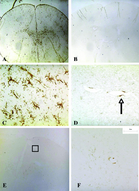
FIG. 1.
Cerebral microgliosis in scid mice persistently infected with serotype 1 of B. turicatae. Immunostaining with rat anti-mouse F4/80 monoclonal antibody shows extensive microgliosis in the brain of a scid mouse persistently infected with serotype 1 of B. turicatae (A). In comparison, little staining is seen in an uninfected control (B) or in a scid mouse persistently infected with serotype 2 (E) (magnification, ×20). Selected areas of the cerebral cortex (shown in squares in panels A, B, and E) are shown at higher magnifications in panels C, D, and F, respectively (×400). The arrow in panel D points to a perivascular F4/80-positive cell.

