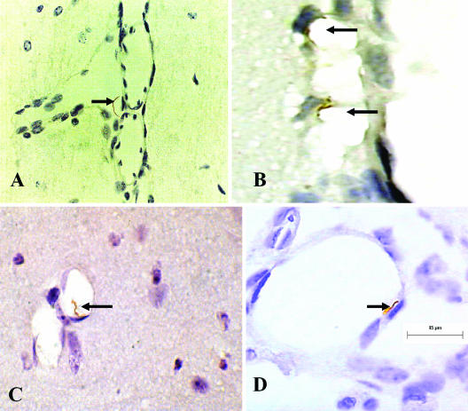
FIG.2.
Interaction of B. turicatae with cerebral microcirculation in vivo. Immunostaining with αVsp1 monoclonal antibody 1H12 of scid mouse brains 1 month after intraperitoneal inoculation with serotype 1 of B. turicatae is shown. Arrows point to spirochetes on the abluminal side of the leptomeningeal microcirculation within the subarachnoid space (A), bound to the luminal side of leptomeningeal cells (B), bound to brain parenchymal endothelial cells (C), and in the process of crossing leptomeningeal endothelial cells (D). 3,3′-Diaminobenzidine chromogen staining; magnification, ×1,000.

