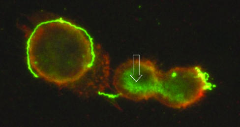
FIG. 3.
Interaction of serotype 1 of B. turicatae with brain microvascular endothelial cells (BMEC) in vitro. BMEC grown on cell culture slides were incubated with serotype 1 spirochetes, washed, incubated with DiI to label BMEC membranes orange, and immunostained with αVsp1 monoclonal antibody 1H12, followed by an FITC-labeled secondary antibody to label Vsp1 green. Microscopic examination with a dual FITC and rhodamine filter revealed green spirochetes next to BMEC. The spirochetes on the surface of BMEC show areas of yellow color, representing colocalization of Vsp1 and BMEC cytoplasmic membrane. Green signal, representing Vsp1, is seen not only extracellularly in spirochetes but also inside BMEC as amorphous material (open white arrow) (magnification, ×1,000).

