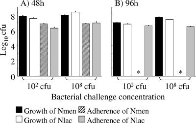
FIG. 6.
Comparison of the growth of Neisseria lactamica (Nlac) and Neisseria meningitidis (Nmen) in culture and association to meningioma cells at time points when cell death is apparent. Monolayers of meningioma cells were infected with various concentrations of N. lactamica and N. meningitidis, and growth in culture and association to cell monolayers were quantified at 48 and 96 h. Data are shown for infection with the low, nonsaturating (102 CFU) concentration or high, saturating (108 CFU) concentration of each bacterium. Infection experiments were carried out in triplicate with two different meningioma cell lines, and data are shown from a typical experiment with meningioma cells from one patient. The columns represent mean log10 bacterial CFU, and the error bars represent the standard deviations from triplicate-infected wells. An asterisk denotes no recovery of N. meningitidis bacteria from monolayers.

