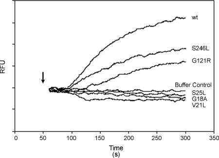
FIG. 7.
Analysis of membrane depolarization induced by mutant VacA proteins. VacA proteins were purified from H. pylori broth culture supernatants as described in Materials and Methods. AZ521 cells were loaded with oxonol VI (a probe used to monitor membrane potential). Following addition of acid-activated VacA proteins (10 μg/ml), changes in fluorescence were monitored. The arrow indicates the time at which toxin was added to the cuvette. An increase in fluorescence (relative fluorescence units [RFU]) over time indicates depolarization of the membrane potential. wt, wild type.

