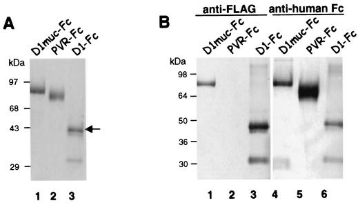
FIG. 2.
Expression of immunoadhesins in CHO cells. (A) The immunoadhesins D1-Fc, D1muc-Fc, and PVR-Fc were purified with protein A columns. The eluted immunoadhesins were analyzed by denaturing SDS-PAGE in 4 to 20% polyacrylamide gels and stained with Coomassie blue. The arrow points to the fully glycosylated form of D1-Fc. (B) Protein A-purified D1muc-Fc (lanes 1 and 4), PVR-Fc (lanes 2 and 5), and D1-Fc (lanes 3 and 6) were analyzed by denaturing SDS-PAGE in a 4 to 20% polyacrylamide gel, transferred to a polyvinylidene difluoride membrane, and probed with anti-FLAG MAb M2 (lanes 1 to 3) or anti-human Fc antibodies (lanes 4 to 6). The positions of prestained molecular size markers and their sizes in kilodaltons are shown.

