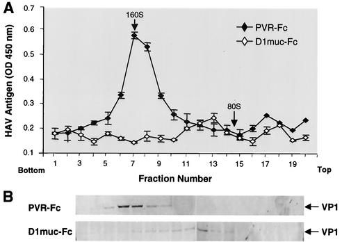
FIG. 6.
Western blot analysis of 100- to 125S HAV particles. (A) Purified cytopathic HAV virions were treated with 10 μg of D1muc-Fc or PVR-Fc and sedimented through 15 to 30% sucrose gradients at 4°C for 90 min with an SW40 rotor at 40,000 rpm. Gradients were collected from the bottom in 20 fractions. HAV antigen in the fractions was determined by capture ELISA. (B) Fractions 4 to 20 of the gradients were incubated with rabbit anti-HAV antibodies and magnetic beads coated with anti-rabbit IgG for 2 h at 4°C. After extensive washing, the beads were boiled in denaturing buffer and proteins were studied by Western blot analysis with staining with guinea pig anti-HAV antibodies and phosphatase-labeled goat anti-guinea pig antibodies. The arrows point to the VP1 structural protein of HAV. The top and bottom of the gradients are indicated. OD, optical density.

