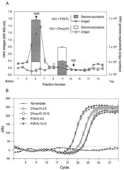
FIG. 7.
Quantification of HAV RNA in 100- to 125S particles. (A) Purified HAV virions were incubated with 10 μg of D1muc-Fc or PVR-Fc for 2 h at 37°C and analyzed through 15 to 30% sucrose gradients as described in the legend to Fig. 6. Fractions containing HAV particles (4 to 6 and 10 to 12) were pooled, and RNA was extracted, ethanol precipitated, and quantitated with a one-step real-time TaqMan RT-PCR assay. In vitro-synthesized full-length HAV RNA was used as the standard for the quantitative PCR. The data shown are mean results from triplicate reactions. Standard deviations are shown as error bars. Poliovirus particles labeled with [35S]methionine were used as 160S and 80S sedimentation markers. The top and bottom of the gradient are indicated. (B) TaqMan amplification plot of each triplicate reaction used to calculate the mean number of HAV genome equivalents in panel A. The ΔRn (increment of fluorescence reporter signal) for each sample was plotted against the cycle number. OD, optical density.

