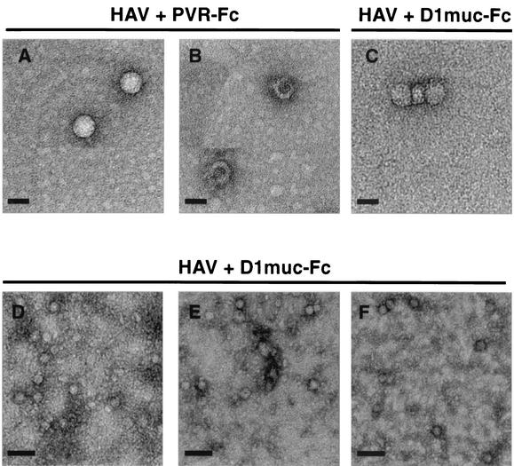
FIG. 8.
Negative-stain EM analysis of HAV particles. HAV was incubated with 10 μg of D1muc-Fc or PVR-Fc for 2 h at 37°C and analyzed through 15 to 30% sucrose gradients as described in the legend to Fig. 6. Fractions containing the 100- to 125S particles from five D1muc-Fc sucrose gradients were pooled and concentrated. Fractions containing 160S and 80S HAV particles from one PVR-Fc sucrose gradient were pooled and concentrated. HAV virions (A), empty capsids (B), and 100- to 125S particles (C) were bound to grids, stained with 2% uranyl acetate, and analyzed by EM at a magnification of ×45,000. Purified KRM003 HAV virions were treated with 30 μg of D1muc-Fc for 10 (D), 30 (E), and 60 (F) min, stained directly without sedimentation, and analyzed by EM at a magnification of ×17,000. Bars: A, B, and C, 30 nm; D, E, and F, 100 nm.

