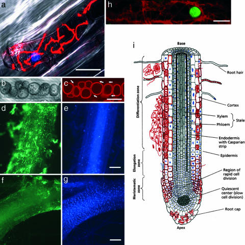
Fig. 3.
Association of fungal structures with living and dead cells of the host tissue. (a) Fungal hyphae swathe a plant protoplast, which undergoes cytoplasmic shrinkage. Hyphae and nucleus stained with WGA-TMR and DAPI, respectively, are superimposed with the bright-field image. (b) Bright-field interference contrast image of chlamydospores in a root cortex cell. (c) Fluorescence image of the same cell stained with fuchsin-lactic acid. Arrows indicate hyphae on which the chlamydospores are formed. (d–g) Root colonization spatially associated with the absence of intact plant nuclei. Root segments (60 hours after inoculation) double-stained for intact plant nuclei (DAPI; e and g) and fungal hyphae (WGA-AF 488; d and f). (d and e) A root segment heavily colonized by fungal hyphae (d) contains only a few DAPI-stained nuclei (e). (f and g) A root segment with minor fungal colonization (f) contains a high number of DAPI-stained nuclei (g). (h) Hyphae swathing a cortical cell protoplast with a TUNEL-positive (green) nucleus. (i) Schematic drawing of a P. indica-infested root showing the different tissues and the associated colonization pattern, with hyphae depicted in red and DAPI-positive plant nuclei depicted in blue. (Scale bars: a, 30 μm; c, 10 μm; d–g, 300 μm; and h, 20 μm.) [Modified from ref. 37 (Copyright 1998, Sinauer, Sunderland, MA).]

