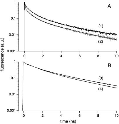
FIGURE 3.
1P time-resolved fluorescence lifetime of diI-C18 and A488-IgE in living mast cells, under different conditions of IgE cross-linking. Representative single-point fluorescence lifetime decays for diI-C18 (A) in the plasma membrane of mast cells in the presence (curve 1) or absence (curve 2) of α-IgE; and A488-IgE (B) on the surface of mast cells with (curve 3) or without (curve 4) cross-linking (λex = 480 nm for both diI-C18 and A488-IgE). Triexponential diI-C18 decays were measured at magic angle polarization and biexponential A488-IgE decays were calculated from the measured parallel and perpendicularly polarized fluorescence decays (following the denominator of Eq. 2). In the calculated magic angle fluorescence decays, the fitting was started beyond the FWHM of the system response function. All decays were fit following Eq. 1, and the fit parameters of time-resolved fluorescence decays are summarized in Table 1. Comparison between both lifetime methods (with and without deconvolution) is included in the text where appropriate. With samples having long excited state lifetimes (i.e., Alexa 488), there are no significant differences in the fitting parameters with each method.

