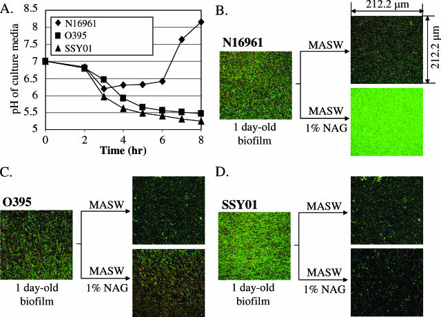
FIG. 7.
Effect of NAG on biofilm viability. (A) pH change of culture supernatants in the NAG-supplemented culture. Culture conditions were identical to those described in the legend for Fig. 1D, except for the use of 1% NAG in the media. (B, C, and D) Confocal laser microscopic analysis of biofilms of N16961, O395, and SSY01. To acquire images, live cells were stained with Syto-9 (green) and dead cells were stained with propidium iodide (red). Top (x-y plane) views were projected from a stack of 25 images taken at 0.5-μm intervals for a total of 12 μm. Before staining, 1-day-old biofilms were treated with MASW (see Materials and Methods) containing 0% (top right panel of each set) or 1% (bottom right panel of each set) NAG for 1 day.

