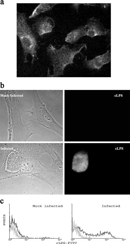
FIG. 1.
Infection of PPEC with C. muridarum. Light and fluorescence micrographs of PPEC. Cell monolayers were infected with C. muridarum (15 IFU/cell) as described in Materials and Methods and examined by fluorescence microscopy 48 h later. (a) PPEC were incubated with anti-cytokeratin antibody and Cy-3-labeled secondary antibodies. (b) PPEC stained with FITC-labeled MAb specific for cLPS. (c) Flow cytometry analysis showing the presence of intracellular cLPS in C. muridarum-infected PPEC. PPEC monolayers were infected with C. muridarum using an MOI of 15 and incubated for 48 h. Cells were treated with 0.1% saponin and incubated with FITC-labeled cLPS-specific antibodies. Figures are representative of at least five experiments performed.

