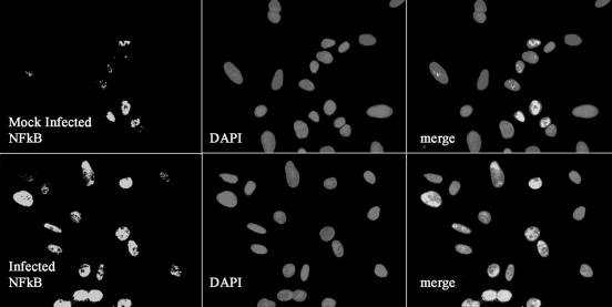
FIG. 4.
NF-κB activation in PPEC induced by C. muridarum infection. PPEC were exposed to C. muridarum at an MOI of 15 for 2 h. Cells were subsequently stained with anti-p65 MAb and Cy-3-labeled secondary antibodies. Nuclei were counterstained with DAPI. NF-κB nuclear localization results in colocalization of red and blue signals resulting in rose fluorescence. Cells were visualized by confocal microscopy. The experiment was performed twice, and the pictures shown correspond to representative fields.

