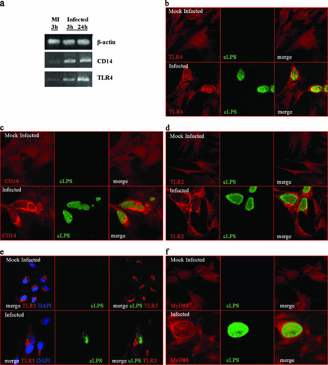
FIG. 5.
TLR4, CD14, TLR2, TLR5, and MyD88 expression in PPEC infected with C. muridarum. (a) RT-PCR analysis of mock-infected or C. muridarum-infected PEC. PPEC were infected at an MOI of 15 with C. muridarum as described in Materials and Methods. Total RNA was extracted at the indicated time after infection and analyzed by a qualitative RT-PCR. Confocal microscopy analysis of TLR4, CD14, TLR2, TLR5, and MyD88 expression on PPEC infected with C. muridarum. PPEC were grown in coverslips and infected with C. muridarum. Forty-eight hours postinfection cells were fixed, permeabilized, and stained using specific antibodies labeled with Alexa 546 or rhodamine. The appearance of chlamydia inclusions was revealed with FITC-labeled MAb specific for cLPS. Fluorescence micrographs of PPEC stained with TLR4-specific antibody (b), with CD14-specific antibody (c), with TLR2-specific antibody (d), and with TLR5-specific antibody. Nuclei were counterstained with DAPI (e) and with MyD88-specific antibody (f).

