FIG. 6.
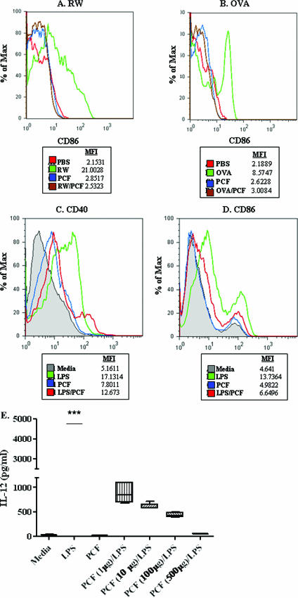
Ex vivo and in vitro analysis of DC activation. CD11c+ cells removed ex vivo from the peritoneal cavity, spleens, and mediastinal LN after either i.p. sensitization with PCF, RW, or RW/PCF (A) or i.p. sensitization with PCF, OVA, or OVA/PCF (B). i.p. injection of either RW or OVA (A and B, respectively; green) led to increased expression of the costimulatory marker CD86 compared to intraperitoneal DC from mice that received sham sensitization with PBS (red) or PCF alone (blue). (A) Intraperitoneal DC derived from RW/PCF-treated mice (brown) showed reduced levels of CD86 compared to DC from mice in the RW alone group (green). (B) Similar reductions were seen in mice given OVA/PCF treatment (brown) compared with injection of OVA alone (green). Expression of LPS-induced CD40 (C) (green) and CD86 (D) (green) was dramatically reduced in BMDC in vitro after preexposure to PCF (D) (blue). (E) LPS-induced production of IL-12 was significantly inhibited in a dose-dependent manner when the BMDC were preexposed with increasing doses of PCF.

