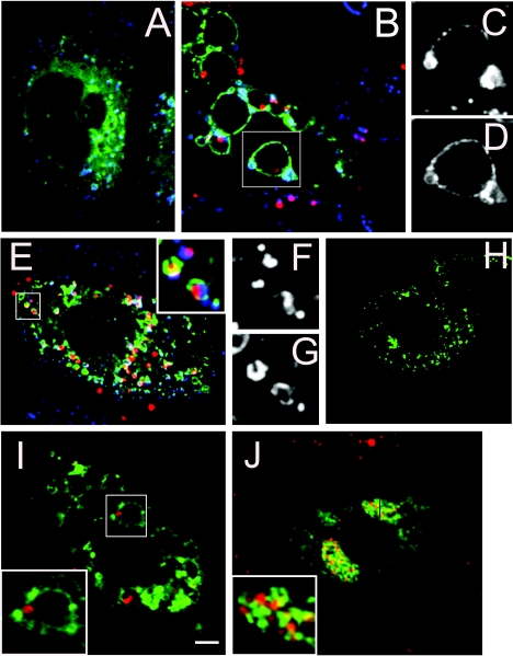
FIG. 2.
Helicobacter pylori's intracellular compartment acquires late endosomal and lysosomal markers. Panels A and B show the distribution of GFP-Rab7 (green) and Lamp-1 (blue) protein for control (A) and wild-type H. pylori-invaded cells (B). Details of the vacuolar compartment showing Lamp1 and Rab7 recruitment are presented in panels C and D, respectively. Panel E shows the distribution of GFP-Rab7 (green) and Lamp-1 (blue) for AGS cells invaded by an H. pylori vacA mutant strain. The inset in panel E shows in detail the morphology of the intracellular compartment of the vacA mutant bacteria. The recruitment of Lamp1 and Rab7 to the bacterial compartment is shown in detail in panels F and G, respectively. Panels H to J show the distribution of GFP-CD63 (green) for uninfected AGS cells (H) and AGS cells infected with wild-type (I) or VacA mutant (J) H. pylori, respectively. The insets in panels I and J show details of the bacterial niches. All of the microphotographs were taken with a spinning disk confocal microscope with a ×100 oil objective. The scale bar in panel I is equivalent to 3 μm. Immunolabeled bacteria are shown in red. For all experiments, the invasion time was 24 h.

