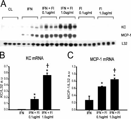
FIG. 3.
IFN-γ priming of skeletal muscle cells augments chemokine expression in response to TLR5 stimulation. (A) RNase protection assay demonstrating chemokine expression by C2C12 myotubes under the following conditions: unstimulated cells (CL), cells primed for 24 h with IFN-γ (IFN; 200 U/ml), cells stimulated for 4 h with flagellin (Fl) alone, and IFN-primed cells stimulated for 4 h with flagellin (IFN + Fl). (B and C) Quantification of chemokine mRNA levels by densitometry, expressed in arbitrary units (a.u.) and normalized to the L32 housekeeping gene (n = 4 per group). *, P < 0.05 versus IFN alone; †, P < 0.05 for comparisons between IFN + Fl 0.01 μg/ml and IFN + Fl 1.0 μg/ml.

