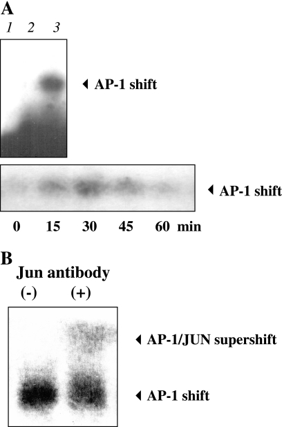
FIG. 4.
Activation of the transcriptional factor activator protein (AP-1) at hBD-3-specific promoter sites in S. aureus-treated keratinocytes as determined by EMSA. Keratinocytes were treated with viable S. aureus and then lysed at various time points for nuclear extract preparation for biotinylated EMSA analysis using oligonucleotide probes based on the AP-1 motifs in the hBD-3 promoter. (A, upper panel) EMSA showing free probe control (lane 1), 100-fold excess unlabeled probe control (lane 2), and AP-1 binding after challenge with S. aureus for 30 min (lane 3). (A, lower panel) Time course of AP-1 binding upon S. aureus stimulation. (B) Supershift of AP-1 complexes binding to the hBD-3 gene promoter is shown in nuclear extracts from S. aureus-treated keratinocytes using polyclonal antibodies to jun family proteins (second lane).

