FIG. 2.
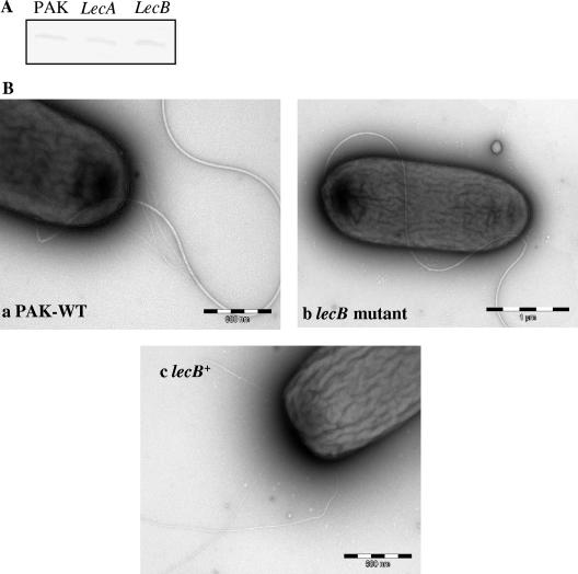
(A) Western blot analysis of PilA production by PAK and by lecA and lecB mutants. PilA was detected with anti-PilA antibody and is indicative of intracellular pilin in these strains. (B) Transmission electron microscopy of PAK, the lecB mutant, and the complemented lecB mutant (lecB+). Cells were used to coat carbon grids, stained with 1% phosphotungstic acid, washed, and dried. The mounted bacteria were viewed with a Hitachi H-7000 transmission electron microscope.

