FIG. 4.
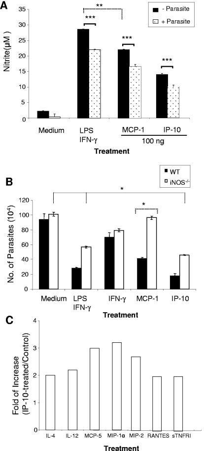
Nitric oxide and proinflammatory chemokines contribute to IP-10- and MCP-1-mediated parasite killing. (A) BM-Mφs of B6 mice (3 × 10⁵ cells/well) in 24-well plates were left untreated or treated with the indicated stimuli, as described in the legend to Fig. 2, for 4 h prior to infection with 2.4 × 10⁶ stationary-phase promastigotes (at an 8:1 parasite-to-cell ratio). At 48 h postinfection, supernatants from uninfected (black bars) and infected (dotted bars) groups were collected for measurement of nitrite via the Griess reagent. (B) BM-Mφs were generated from wild-type B6 mice (black bars) or iNOS−/− B6 mice (white bars) and treated with 100 ng/ml of IP-10, MCP-1, or IFN-γ for 4 h prior to infection with L. amazonensis lesion-derived amastigotes as described in the legend to Fig. 2. Cells treated with LPS (20 ng/ml) plus IFN-γ (20 ng/ml) served as positive controls. At 48 h postinfection, cells were treated with 0.01% SDS to release intracellular parasites, and the parasite number per well was counted. Data are presented as numbers of parasites per well and expressed as means ± SD for triplicate wells per condition. The iNOS−/− groups were compared with their wild-type counterparts ( , P < 0.05;
 , P < 0.01);

 , P < 0.001). The data shown are representative of three independent repeats. (C) BM-Mφs of BALB/c mice were seeded into six-well plates (1 × 10⁶ cells/well) and infected with 8 × 10⁶ stationary-phase promastigotes (8:1 parasite-to-cell ratio). At 48 h postinfection, cell-free supernatants were collected for the measurement of cytokine profiles via protein cytokine arrays. The intensities of protein spots for the IP-10-treated group were compared with those of the corresponding spots for the untreated controls, and data are presented as x-fold increases above the infection control levels. The data shown are the results for those molecules that displayed ≥2-fold increases over the infection control levels and are representative of three independent repeats.

