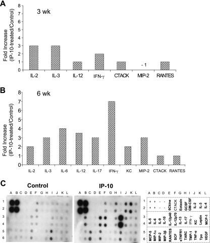
FIG. 6.
Local administration of IP-10 triggers the production of multiple Th1-favored cytokines and chemokines. B6 mice (five per group) were infected and treated as described in the legend to Fig. 5. At 3 weeks (A) and 6 weeks (B) postinfection, LN cells (5 × 10⁶/ml/well) were collected from IP-10-treated groups or PBS controls, pooled from five mice, and stimulated with parasite antigen (1 × 10⁷ parasite equivalents) for 48 h. Cell-free supernatants were collected for the measurement of cytokine profiles via protein cytokine arrays. The intensities of protein spots for the IP-10-treated group were compared with those of the corresponding spots for the infection controls, and data are presented as x-fold increases above the infection control levels. Data shown in panels A and B represent those molecules with ≥2-fold increases compared to the infection control levels at 3 and 6 weeks postinfection, respectively. Example membranes for infection control and IP-10 groups at 6 weeks postinfection are shown in panel C, and all tested cytokines/chemokines in the membranes are illustrated to the right. Membranes for medium controls using cells from infected mice detected no or minimal expression of cytokines and chemokines (data not shown).

