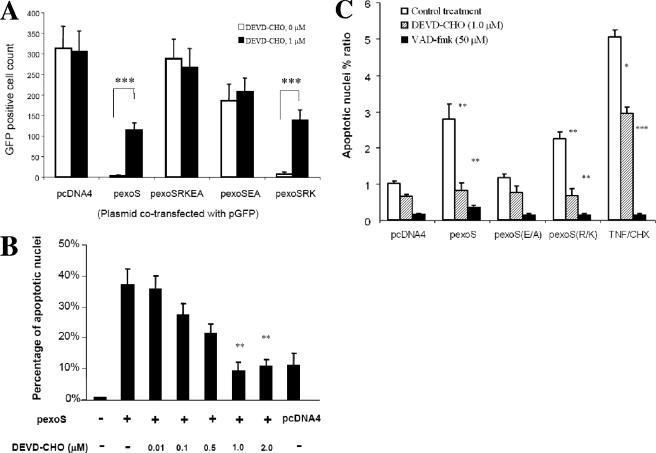
FIG. 1.
Caspase-3-dependent cytotoxicity caused by transfection of the exoS expression plasmid. (A) Disappearance of GFP signal after cotransfection. HeLa cells were cotransfected with pGFP reporter plasmid and the exoS expression plasmids as indicated, with (▪) or without (□) the presence of 1 μM DEVD-CHO, a cell-permeable caspase-3 inhibitor. HeLa cells were observed under a fluorescence microscope using GFP fluorescence at 24 h posttransfection, and GFP-positive cells were quantified from five random views. Plasmids used for cotransfection with pGFP are indicated as follows: pcDNA4, vector control; pexoS, wild-type exoS in pcDNA4(pJJ0040); pexoSEA, exoS(E381A) in pcDNA4(pJJ0043); pexoSRK, exoS(R146K) in pcDNA4(pJJ0042); and pexoSRKEA, exoS(R146K/E381A) in pcDNA4(pJJ0044). As a solvent control, HeLa cells were treated with dimethyl sulfoxide (DEVD-CHO, 0 M). The data are means ± the SD of the counts of GFP-positive cells from three replicates. P values were calculated by comparing DEVD-CHO-treated (1 μM) and untreated (0 M) groups (***, P < 0.001). (B) Nuclear condensation caused by transfection with pcDNA4-exoS. HeLa cells were transfected with pexoS, wild-type exoS in pcDNA4, or pcDNA4 alone. HeLa cells were collected at 36 h posttransfection, stained with Hoechst dye, and subjected to fluorescence microscopy. Five fields were randomly sampled from each experimental population, and all of the cells stained with Hoechst dye in each field were counted up to 500 in total. The total number of apoptotic cells with condensed or fragmented nuclei was determined in the five sampled regions and was expressed as follows: percentage of apoptosis per sample = (number of apoptotic cells/total number of cells) × 100%. During and after transfection, HeLa cells were treated with the indicated amount of DEVD-CHO, a cell-permeable specific caspase-3 inhibitor. The data are means ± the SD of the percentages from three replicates. Significant differences between certain DEVD-CHO treated and untreated groups are indicated (**, P < 0.01). (C) Apoptosis after transfection with the indicated exoS expression constructs. In each experiment, the apoptotic nucleus percent ratio was calculated by comparing the calculated percentage of apoptosis to that of the pcDNA4. During and after transfection, HeLa cells were treated without (control) or with indicated amounts of DEVD-CHO or VAD-fmk, a cell-permeable specific caspase-3 or a pan-caspase inhibitor, respectively. TNF/CHX, treatment with TNF-α and CHX was used as a positive control for apoptosis induction. The data are means ± the SD of the ratios from three replicates. Significant differences between certain DEVD-CHO- or VAD-fmk-treated and untreated control groups are indicated (*, P < 0.05; **, P < 0.01; ***, P < 0.001).

