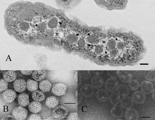
FIG. 1.
Carboxysomes of H. neapolitanus. (A) Transmission electron micrograph of an H. neapolitanus cell containing carboxysomes. (B) Purified, negatively stained intact carboxysomes. (C) Negatively stained carboxysomes after rupture by freeze-thaw treatment. In all panels, the bar represents 100 nm.

