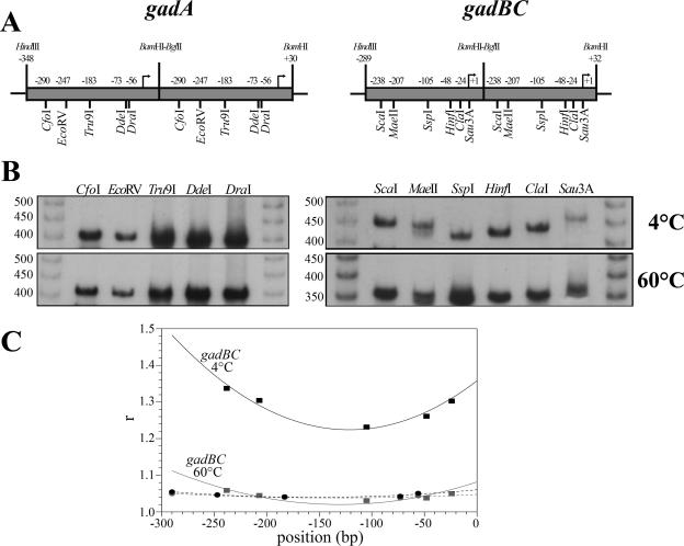
FIG. 5.
Gel electrophoretic mobility assay of the circularly permutated gadA and gadB fragments. (A) Maps of the gadA and gadB head-to tail dimers. The positions of single cutting restriction sites are indicated. The bent arrows indicate the transcriptional start sites. (B) Gel electrophoresis of the radiolabeled gadA and gadB permutated fragments, performed with a 5% polyacrylamide gel at 4°C (upper panel) and 60°C (lower panel). The sizes of members of a nonbent 50-bp ladder are indicated on the left. (C) Ratios apparent size to real size (r) of gadA (•) and gadB (▪) permutated fragments plotted against map positions (bp). The dashed (gadA) and solid (gadB) lines are the lines of fit to a parabolic function.

