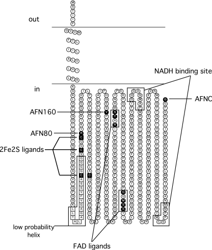
FIG. 7.
Membrane topology model of NqrF. The binding sites for NADH, the 2Fe-2S center, and FAD are indicated. The residues of the weak hydrophobic region predicted to be a possible second transmembrane helix are shown as white squares. C-terminal fusion sites are indicated in the figure using the codes shown in Table 1.

