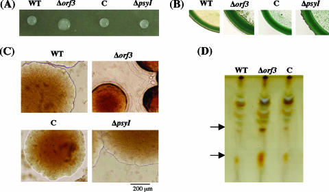
FIG. 3.
Detection of biosurfactants, colony morphology, and separation of glycolipids. (A) Drop-collapsing test of bacterial suspension. (B) Methylene blue plate assay. (C) Colony morphology of the WT and each mutant observed by optical microscopy on 1.5% KB agar plate after 2 days of incubation at 27°C. (D) TLC analysis of glycolipids. Arrows indicate orcinol-positive spots with higher intensity in the Δorf3 mutant. Bacterial strains are indicated as: C, orf3-complemented strain.

