
Fig. 2.
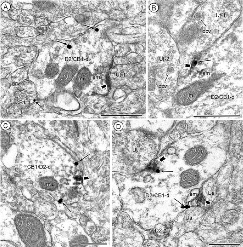
Dendritic distributions of D2 and CB1 receptors. Acb core (A), shell (B) processed for D2-peroxidase and CB1-gold immunocytochemistry. Dually labeled dendrites (D2/CB1-d) show peroxidase reaction product (block arrow) on the plasma membrane beneath unlabeled terminals (Ut-1). Gold-silver particles (small arrows) are located either distant (A) or within (B) the peroxidase aggregates at the dendritic surface. In A, gold labeling (small arrow) is also located on the plasma membrane of a nearby axon (CB1-a). Both the CB1-labeled axon in A and the unlabeled axon terminals (Ut-1 and Ut-2) in B contain numerous large dense core vesicles (dcv). C: Acb shell, reversal of labels, CB1-immunoperoxidase and D2-immunogold. Both the gold-silver (small arrows) and peroxidase product (block arrow) are located on or near the plasma membrane in a common dendrite (CB1/D2-d). D: Acb shell processed for D2-peroxidase and CB1-gold immunocytochemistry. Aggregates of peroxidase (block arrow) and gold particles (small arrows) show overlapping distributions near endomembranes in a dually labeled dendrite (D2/CB1-d). The labeling is prevalent near regions contacted by unlabeled axons (ua) or an unlabeled terminal (Ut). Peroxidase D2-labeling is also seen in a nearby small axon (D2-a). m, mitochondria; em, endomembranes. Scale bars = 0.5 μm.