
Fig. 4.
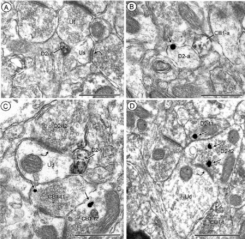
Separate localization of D2 and CB1 receptors in axonal profiles. A: Acb core, processed for D2-peroxidase and CB1-gold. The peroxidase reaction product is located on the plasma membrane and throughout the cytoplasm in a small unmyelinated axon (D2-a) apposing an unlabeled terminal (Ut) and an unlabeled dendritic spine (Us). This spine receives an asymmetric synapse (curved arrow) from an unlabeled axon terminal. B: Acb core, reverse markers with CB1-peroxidase and D2-gold. Two adjacent small axons show plasmale-mmal immunogold (small arrow; D2-a) or diffuse peroxidase reaction product (CB1-a). C: Acb shell, processed for D2-peroxidase and CB1-gold. D2 immunoreactivity is distributed throughout a small preter-minal axonal process (D2-t1) and on the apposed plasma membrane and subsurface vesicles within a large terminal (D2-t2), which forms an asymmetric synapse (curved arrow) with an unlabeled dendrite (Ud). A convergent terminal (CB1-t1) on the same dendrite and another terminal (CB1-t2) within the neuropil contain isolated immu-nogold particles (small arrows) indicating the presence of CB1 receptors. D: Acb shell, with reversal of markers, CB1 peroxidase and D2-gold. Immunogold (small arrows) labeling is seen in two apposed terminals (D2-t1 and t2), the latter of which forms what appears to be a tangentially sectioned asymmetric synapse (curved arrow) with an unlabeled dendrite (Ud). The dendrite is apposed by an axon terminal showing diffuse CB1-immunoperoxidase labeling (CB1-t). Scale bars = 0.5 μm.