
Fig. 8.
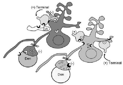
Schematic diagram showing co-expression and partially intracellular distributions of CB1 (white block arc) and D2 (black block arc) in a spiny neuron (light gray shading), whose local inhibitory (−) terminal also contains both markers. An adjacent spiny neuron (dark gray) expresses CB1 (white block arc), but not D2 receptors. Putative dopaminergic (DA) and apposed excitatory (+) type terminals, both containing D2 receptors, are presynaptic to the dendrite that contains CB1 receptor. Conversely, the dendritic profiles (light gray shading) labeled for D2 receptors including those that also contain CB1 receptors are postsynaptic to CB1- or CB1- and D2-labeled excitatory inputs (+). Inhibitory-type (−) terminals, likely originating as collaterals of the spiny projection neurons (double bar), are immunolabeled for either CB1 or CB1 and D2. These are shown as presyn-aptic to dendrites (Den) that are unlabeled (white stipple) or labeled for the D2 receptor.