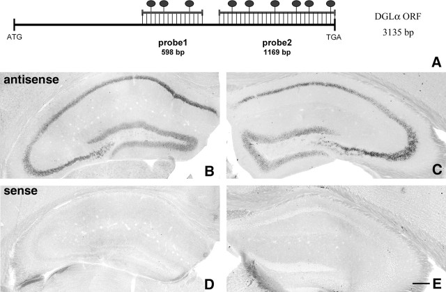
Figure 1.
Principal cells express high levels of DGL-α mRNA in the hippocampus. A, Schematic representation of the positions of the two complementary antisense riboprobes on the sequence of the mouse DGL-α mRNA. The length of the open reading frame (ORF) on the DGL-α mRNA is 3135 bp long. The ATG codon indicates the translation initiation site on the sequence and represents position 1–3 in the numbering, whereas TGA is the stop codon (position 3133–3135). The dark ovals above the duplex scheme indicate the predicted frequency of digoxigenin-labeled nucleotides in the ratio of the total length of the probes, which were 598 and 1169 bp in the case of probe 1 and probe 2, respectively. B, C, In situ hybridization by using the two antisense probes illustrated in A visualizes the principal cell layers of the mouse hippocampus. The expression level is very high in the CA1 and CA3 pyramidal neurons and somewhat weaker, but still high, in the granule cells of the dentate gyrus. In contrast, neither GABAergic interneurons nor glial cells are labeled by the probes under these reaction conditions, indicating much weaker expression or a complete absence of DGL-α in these cell types. Note the identical labeling by the two riboprobes, which confirms the specificity of the signal. D, E, In contrast, in situ hybridization using the sense riboprobes derived from the corresponding DGL-α sequence do not result in any labeling. Scale bar, 200 μm (for B–E).

