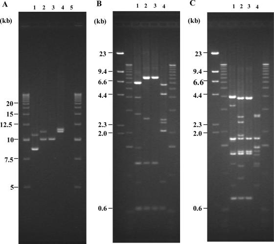
FIG. 2.
(A) Agarose gel electrophoresis patterns of PCR amplicons obtained against region V of Stx phage. Lanes: 1, I-A (RIMD0509952, original pattern); 2, II-A (EC1015, original pattern); 3, II-B (EC1015, lost Stx2 phage pattern); 4, III-A (83-1386, original pattern); 5, C600. Ladders (2.5 kb) were used as molecular mass markers on both sides of the gel. (B and C) BglI digest (B) and EcoRV digest (C) of LA-PCR products obtained from region V in STEC O157:H7 strains RIMD0509952, EC1015, and 83-1386. Lanes: 1, I-A (RIMD0509952, original pattern); 2, II-A (EC1015, original pattern); 3, II-B (EC1015, lost Stx2 phage pattern); 4, III-A (83-1386, original pattern). Lambda-HindIII digest and 1-kb ladders were used as molecular mass markers on both sides of each gel. Positions of DNA fragments with known molecular masses are indicated.

