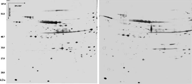
FIG. 2.
Two-dimensional gel of T. whipplei extract with silver staining (the first one is for Slow2, the second one is for Endo5). Proteins were resolved in the first dimension over a pI gradient of 4.5 to 5.5, followed by a second-dimension separation by SDS-PAGE in a 10% acrylamide gel. The prominent spots at 60 kDa and 84 kDa (arrow) were cored from the gel and submitted for analysis by mass spectrometry. These spots corresponded to the 2-DE blotting.

