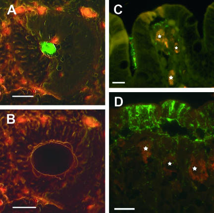
FIG. 1.
In situ visualization of the novel “Ca. Treponema suis” colonizing colonic tissue samples of pigs. The bacteria were visualized with green fluorescent 16S rRNA oligonucleotide probes, and the intestinal cell material exhibited red autofluorescence. (A and B) Cross-section of a colonic crypt before and after microdissection, respectively. The bacterial cells were visualized with a probe targeting spirochete species (S-G-Leptospira-1414-a-A-18). Scale bar, 50 μm. (C) Fluorescent in situ hybridization with a probe specific for “Ca. Treponema suis” (S-S-Treponema-0833-a-A-18) demonstrating a cluster of “Ca. Treponema suis” in a crypt and single spirochetes within lamina propria surrounded by large macrophages (stars). Bar, 15 μm. (D) Porcine colonic mucosa with severe focal spirochetal infiltration by “Ca. Treponema suis” (S-S-Treponema-0833-a-A-18). High numbers of the spirochete are demonstrated within the surface epithelium and adjacent lamina propria, while colonization of the surface is absent. Large macrophages are indicated by stars. Bar, 20 μm.

