
Fig. 6.
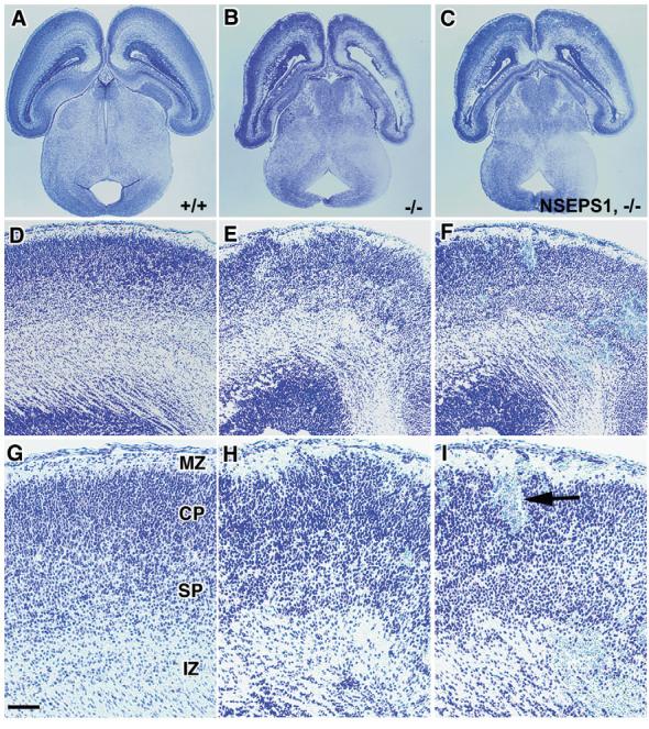
Failure of an NSE-Psen1 transgene to rescue the brain pathology in Psen1-/- mice. Shown are Cresyl Violet-stained horizontal sections of E18.5 embryos that are wild type (A,D,G), Psen1-/- (B,E,H) or have the NSE-Psen1 transgene on Psen1-/- background (C,F,I). (D-I) Higher power views of the lateral cerebral wall. The embryo with the NSE-Psen1 transgene on the Psen1-/- background exhibits multiple areas of hemorrhage (one indicated by arrow), as well as disrupted cortical lamination in a pattern that is indistinguishable from that of the Psen1-/-embryo. Cortical layers (MZ, CP, SP, IZ) are indicated as in Fig. 5. Scale bar: 500 μm in A-C; 100 μm in D-F; 50 μm in G-I.