
Fig. 9.
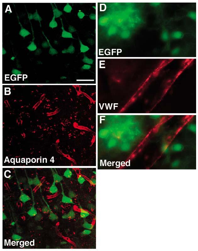
Lack of the nestin intron 2 enhancer activity in vascular progenitor cells or endothelial cells. NesCrenls line 27 was crossed with a Cre/loxP reporter line cActXstopXEGFP44. The hippocampal CA1 region of an adult double transgenic mouse is shown in A-C. Spontaneous EGFP fluorescence (A) was imaged and combined with immunohistochemistry for aquaporin 4 (B). EGFP is prominently expressed in the pyramidal cells but not in the aquaporin 4 outlined vessels (merged image is shown in C). (D-F) Combined immunohistochemical staining is shown for EGFP (D), and von Willebrand factor (E) on an adult brain from a NesCrenls line 27 mouse crossed to the Z/EG reporter line (merged image is shown in F). Immunostaining in D-F was performed on sections cut from tissue perfusion fixed with paraformaldehyde. A large penetrating vessel is visible at the cortical surface. There is no EGFP labeling of the von Willebrand factor-stained vessel. Scale bar: 50 μm for A-C; 10 μm for D-F.