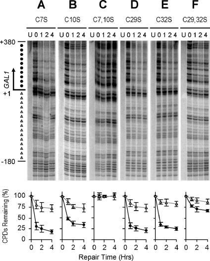
FIG. 4.
TCR mediated by mutated Rpb9. Gels show TCR in the GAL1 gene of log-phase rad16 rad26 rpb9 cells transformed with plasmids encoding Rpb9 with cysteine 7, 10, 29, and 32 replaced by serine. Lanes labeled U represent unirradiated samples. Lanes 0, 1, 2, and 4 indicate different times (hours) of repair incubation following UV irradiation. An arrow to the left of the gels marks the transcription start site. Solid circles on the left of the gels mark transcribed region (+1 to +380). Open triangles mark the upstream region (−1 to −180) where residual Rpb9-mediated TCR takes place. Plots underneath each of the gels show the average (mean ± standard deviation) of the percent CPDs remaining at individual sites in the transcribed (+1 to +380) (solid circles) and upstream (−1 to −180) (open triangles) regions at different times of repair incubation.

