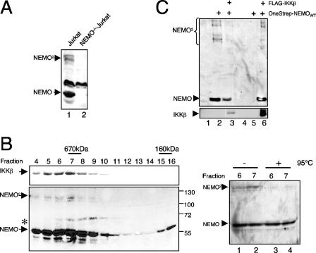
FIG. 4.
Endogenous NEMO dimers are recruited to the IKK complex. (A) Dimer formation of endogenous NEMO. Fifty micrograms of whole-cell extract from wild-type Jurkat T cells (lane 1) or a NEMO-deficient Jurkat mutant (lane 2) was subjected to an anti-NEMO immunoblot analysis. (B) NEMO dimers comigrate with the IKK complex. S100 extracts from 4 × 10⁸ HeLa cells were fractionated on a Superose 6 column, and 6% of the indicated fractions was subjected to an anti-IKKβ (upper panel) or an anti-NEMO (lower panel) immunoblot analysis. The asterisk marks a nonspecific signal. As a control, 6% of the indicated fractions were either subjected to an additional heating step (right part, lanes 3 and 4) or was left untreated (lanes 1 and 2) prior to an anti-NEMO immunoblot analysis. (C) IKKβ interacts with NEMO monomers and dimers. 293 HEK cells were transiently transfected with 2 μg of expression vectors for FLAG-IKKβ and OneStrep-NEMO, as indicated, and the resulting whole-cell extracts were used for an anti-FLAG immunoprecipitation and subsequent anti-NEMO (upper part) or anti-IKKβ (lower part) immunoblot analysis.

