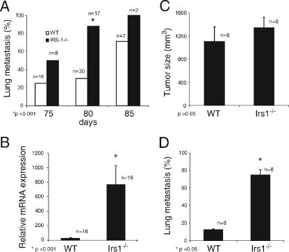
FIG. 3.
Analysis of Irs-1 involvement in PyV-MT mammary tumor metastasis. (A) Lungs from female PyV-MT and PyV-MT::Irs1−/− mice were sectioned and screened microscopically for the presence of metastatic lesions. Five representative sections from each lung were analyzed. The percentage of mice that scored positively for metastatic lesions at each time point (75, 80, and 85 days) is shown (for WT versus Irs1−/− results at 80 days, P < 0.001). (B) PyV-MT mRNA was amplified and quantified from the lungs of 80-day-old PyV-MT and PyV-MT::Irs1−/− mice by RQ-PCR. The data shown are the mean mRNA expression levels (± SEM) from 16 mice of each genotype. (P < 0.001.) (C) Mean tumor burden (± SEM) of PyV-MT and PyV-MT::Irs1−/− tumors grown orthotopically in the mammary fat pads of female nu/nu mice. The number of mice analyzed is indicated. (P > 0.05.) (D) Lungs from mice with PyV-MT and PyV-MT::Irs1−/− orthotopic tumors were sectioned and screened microscopically for the presence of metastatic lesions. Five representative H&E sections from each lung were analyzed. The percentage of the mice that scored positively for metastatic lesions is shown. (P < 0.05.) WT, PyV-MT mice; Irs1−/−, PyV-MT::Irs1−/− mice.

