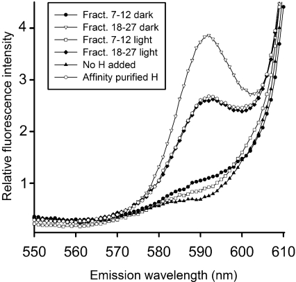
Figure 5. Magnesium chelatase activity of gel filtration fractions of light- and dark-exposed His-tagged H-subunit.
The amount of formed Mg-protoporphyrin IX was analysed by spectrofluorimetry using a λex of 418 nm. Mg-protoporphyrin IX forms a peak at 590 nm. Light- and dark-treated H-subunits from pooled fractions 7–12 and 18–27 in Figure 2(A) were used in the analysis.

