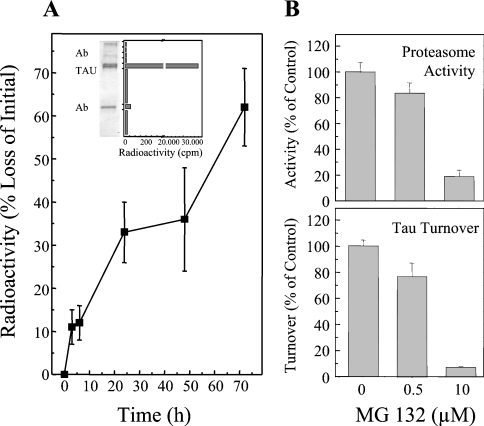
Figure 1. Measurement of tau turnover by the proteasome in HT22 neuronal cells.
Cells were labelled with [3H]lysine for 72 h. Tau was then immunoprecipitated and analysed by electrophoresis. To measure the radioactivity in the tau proteins the resulting gel was cut into bands and the bands were analysed by scintillation counting. The immunoblot inset in (A) shows that such a radioactivity profile contains one major band which is the tau protein as revealed by immunoblotting. Turnover of the tau protein was measured by this technique, and the results are presented in the main part of (A). (B) The inhibition of proteasome activity, as measured by cleavage of the suc-LLVY-AMC peptide substrate, by MG132. (C) Inhibition of cellular tau degradation by the proteasome determinations.

