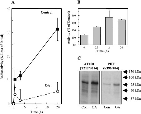
Figure 4. Tau hyperphosphorylation and decreased turnover after treatment of HT22 neuronal cells with OA.
(A) HT22 cells were labelled with [3H]lysine for 72 h, and then treated for 3 h with 0.5 μM OA (or used as controls). Tau was then immunoprecipitated and analysed for radioactivity to calculate tau degradation, as described in Figure 1. (B) Cell lysates were tested for proteasome activity before, and at various time points after, OA treatment, using degradation of the fluorogenic substrate suc-LLVY-MCA as described in the Materials and methods section. The data in both (A) and (B) represent the means±S.E.M. for three independent determinations. (C) Immunoreactivity of HT22 cell lysates to the AT100 and PHF-1 (hyperphosphorylated tau) antibodies in control cells, and after treatment with OA.

