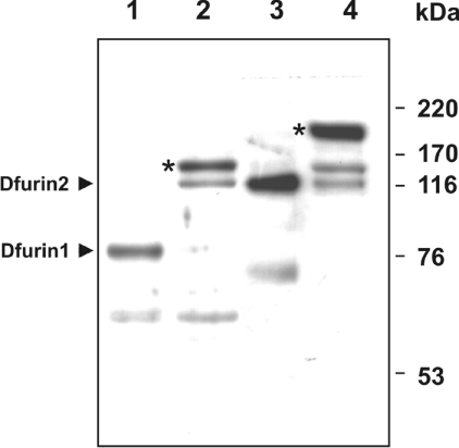
Figure 2. Formation of SDS-stable complexes between recombinant Spn4E and Dfurin1 or Dfurin2.
Supernatants (20 μl) from S2 cells were incubated in the absence (lanes 1 and 3) or presence (lanes 2 and 4) of 50 ng of recombinant Spn4E. After reducing SDS/PAGE (10% gels), the reaction products were analysed by Western blotting, using antibodies directed against Dfurin1 (lanes 1 and 2) or Dfurin2 (lanes 3 and 4) respectively. An asterisk marks Spn4E–Dfurin1 and Spn4E–Dfurin2 complexes depicting the expected sizes. The molecular masses of marker proteins are indicated on the right.

