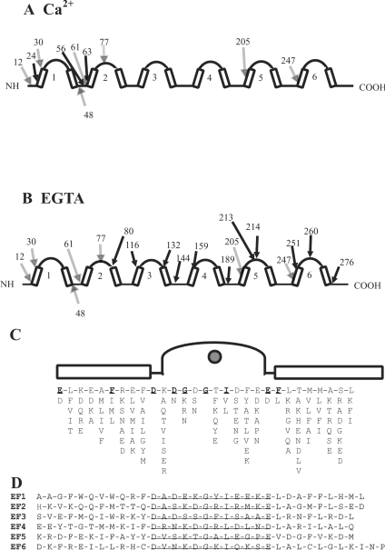
Figure 3. Secretogogin primary and secondary structure.
(A) Limited proteolysis of secretagogin. Schematic diagram of secretagogin outlining secondary structure elements, including the helix–loop–helix motifs and major tryptic cleavage sites in (A) 1 mM CaCl2 and (B) 1 mM EGTA. The residue number of cleavage sites identified both in CaCl2 and EGTA are indicated by shaded arrows. Cleavage sites identified only in CaCl2 or EGTA are indicated by solid arrows. EF-hand motifs 1–6 are shown by arabic numbers. (C) The EF-hand consensus sequence. (D) The amino acid sequence of the six EF-hands of secretagogin. Loops are underlined.

