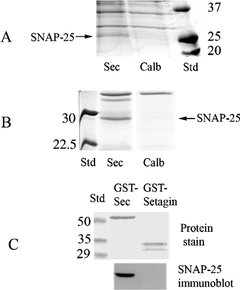
Figure 6. Identification of secretagogin-binding proteins.
Mouse brain tissue (A) and bovine brain tissue (B) were homogenized as described in the Experimental section and incubated with secretagogin (Sec)–Sepharose or calbindin D28k (‘Calb’)–Sepharose respectively for 2 h. The affinity columns were then rinsed followed by elution with a buffer containing 2 mM EGTA. The eluates from all experiments were concentrated by ultra-filtration to 50 μl, and 10 μl was separated by SDS/PAGE. Bands indicated with arrows were cut and processed for MS/MS (gel slice named ‘Sec’). (C) GST–secretagogin or GST–setagin was incubated with cell extracts from RIN-5F cells and pulled down using glutathione beads. After washing the beads, they were boiled in SDS/PAGE sample buffer, centrifuged and transferred on to PVDF membranes and stained or probed using anti-SNAP-25 antibodies.

