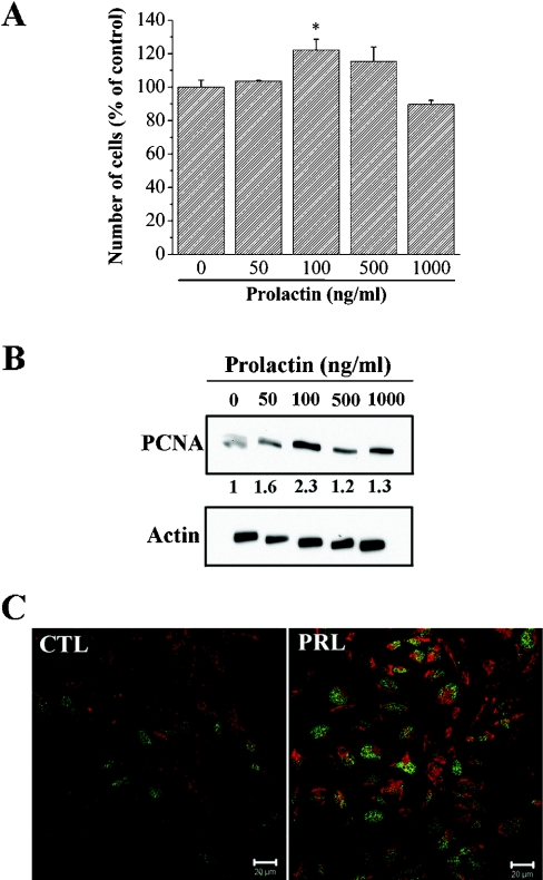
Figure 1. PRL stimulates cell growth.
(A) Cells were seeded in 96-well plates containing 200 μl culture medium with 1% (v/v) fetal calf serum. After 48 h, the cells were treated for 4 days with the indicated dose of PRL. The medium was replaced every 48 h. After 4 days, the number of cells in each well was determined by the MTS assay. (B) Western blot analysis of PCNA expression in cells treated with 100 ng/ml PRL for 24 h. (C) Regulation of PCNA protein expression was investigated by immunofluorescence. PCNA proteins were detected in basal conditions (CTL) and in cells treated with 100 ng/ml PRL for 24 h (PRL). PCNA, green; Evans Blue, red; scale bar, 20 μM. * Significantly different from the control value (P<0.05).

