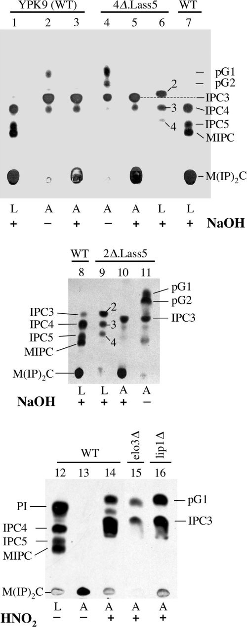
Figure 6. GPI anchor lipids of WT and mutant 4Δ.Lass5 cells are similar.
Labelling of 25 A600 units of YPK9 (WT), 4Δ.Lass5, 2Δ.Lass5, 4Δ.Lass5 elo3Δ (lane 15) and 4Δ.Lass5 lip1Δ (lane 16) cells for 2 h with 100 μCi of [3H]inositol. Free lipids were extracted, GPI proteins were completely delipidated and their lipid moieties were liberated using HNO2, with the exception of lane 13. Free lipids (L, lanes 1, 6–9 and 12) and liberated anchor lipids (A, lanes 2–5, 10, 11 and 13–16) were treated with NaOH (+) or mock incubated (−) for deacylation, separated by TLC and exposed for fluorography. IPCs were annotated as in Figure 3(A).

