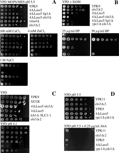
Figure 7. Phenotypic analysis of 4Δ.Lass5 cells under various stress conditions.
Serial 10-fold dilutions of cells were plated on buffered (A and D) or unbuffered YPD + uracil + adenine plates containing the indicated ingredients. Plates were then incubated at 30 °C and photographed after 3 days except for panel (D), where they were grown at 24 °C for 3 days. 4Δ.LAG1 cells were grown in YPG before being plated on YPD, where they strongly reduce sphingolipid biosynthesis but, nevertheless, continue to grow for several days. The vma4Δ cells served as a positive control and exhibited the same Vma− phenotype as described for vma2Δ and vma13Δ [23].

