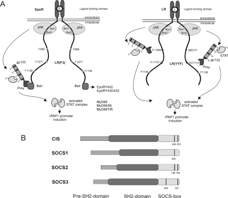
Figure 1. MAPPIT.
(A) MAPPIT, a cytokine receptor-based two-hybrid method, is displayed in the left-hand panel, with the various receptor motifs used in the present study. The right-hand panel shows a variant of the MAPPIT technique using the STAT3 signalling-deficient LR as bait. Both MAPPIT methods are described in more detail in the Results section. (B) Schematic structure of SOCS proteins. Conserved tyrosine residues in the SOCS-box are indicated with a black line.

