Figure 1. .
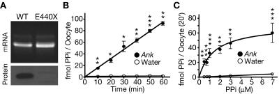
ANK stimulation of PPi translocation across the plasma membrane. A, Agarose gel of wild-type (WT) Ank and mutant ank cRNAs, with western blot of membrane-enriched protein lysates from Xenopus oocytes 3 d after cRNA injection. The cRNA appears intact, and the ANK protein was detected. The mutant ank cRNA encodes a truncated protein (E440X) that does not contain the C-terminal epitope recognized by the ANK antibody and therefore served as a control for antibody specificity. B, Time course of PPi uptake by oocytes in transport buffer with 1 μM 33PPi. After subtracting uptake by water-injected controls, the calculated rate of ANK-dependent transport is 1.54±0.04 fmol per oocyte per min (n=3). C, Rate of ANK-stimulated PPi transport as a function of substrate concentration. The transport rate was saturable, and the apparent Km was 1.33±0.59 μM after subtracting uptake by water-injected controls (n=8). A single asterisk (*) indicates P<.05; a double asterisk (**) indicates P<.01; a triple asterisk (***) indicates P<.001.

